1 .
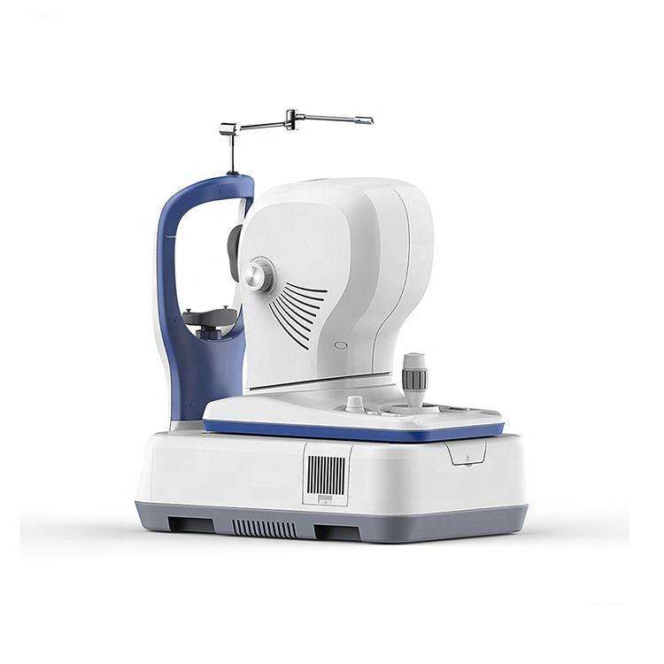
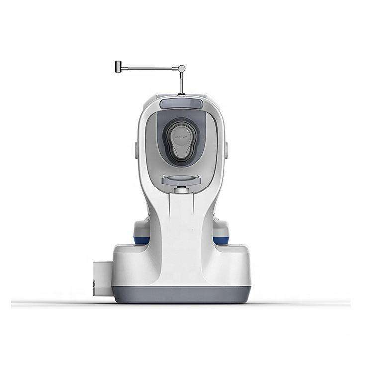

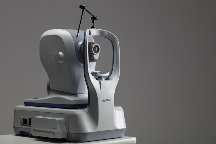
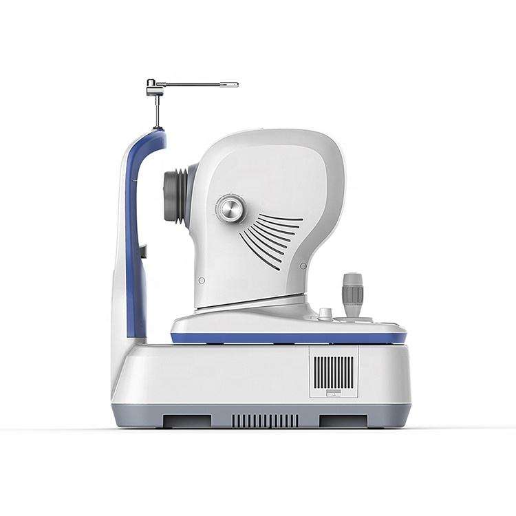
Ophthalmic OCT Machine Optical Coherence Tomography OSE-2800 for Anterior and Posterior Segment of eye
OSE2800 anterior and posterior segment
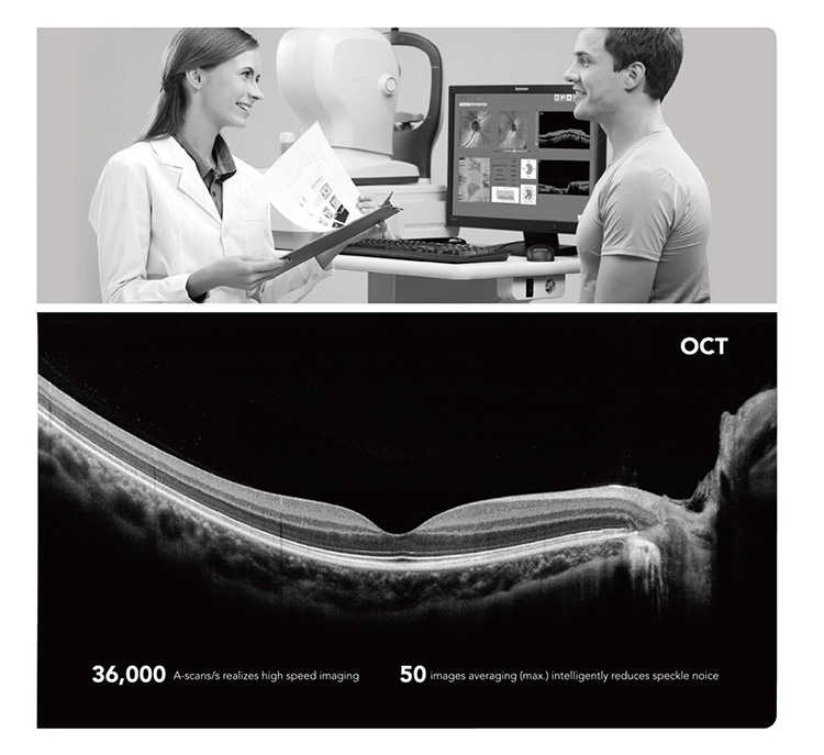
Optical coherence tomography (OCT) is a new , noninvasive , noncontact , transpupillary imaging,technology which can image retinal structures in vivo with a resolution of 5-8 microns. Cross-sectional images of the retina are produced using the optical backsattering of light in a fashion analogous to B-Scan ultrasonography and cofocal microscopy. Cross-sectional images of the retina, is revolutionizing the early detection and treatment and greatly enhanced our quality of patient care.
Applications
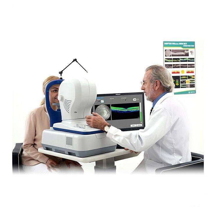
In vivo,cross-sectional images and quantitative analysis of retinal fratures to optimize the diagosis and monitoring of retinal disease and for enhanced pre-and post-therapy assessment.
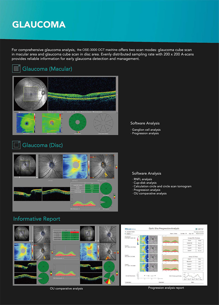
High-quality images and accurate measures RNFL and the optic nerve head to aid in the detection and management of glaucoma.
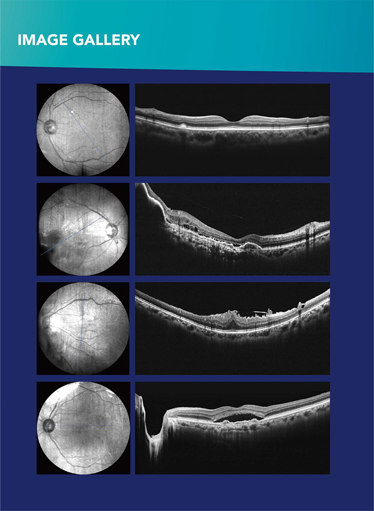
Cross-sectional images are valuable for clinical evaluation of macular holes, macular edema and other retinal pathologies. Precise location of pathology to expand disgnostic confidence and therapeutic precision.
Normal and abnormal image contrast image
Lesions image by image with normal OCT images, the regional thickness values? Topographic maps, diagrams and other multi-thickness contrast, thereby comprehensive judgment of disease.
High performance at a low price.
Modular design increases flexibility , reusability and maintainability.We can provide personalized design according to the customers needs.
With the powerful software , Omhas clear , easy-to-ure interface and supports multi-language.
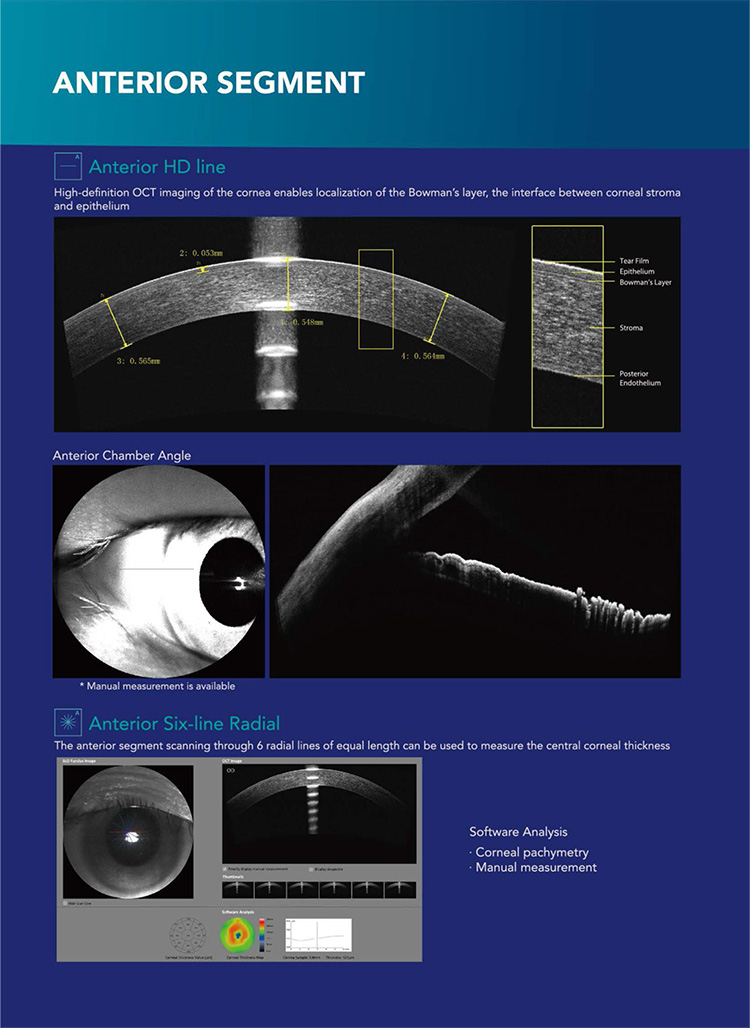
Anterior segment analysis template
Anterior segment with optional modules, OSE 2800 be able to observe and analyze the anterior segment.
Variety of color display , personalized to meet different doctors Figure habits.
| OCT IMAGING | ||
| Methodology | Spectral Domain Oct | |
| Optical Source | Super luminescent diode(SLD),840nm | |
| Scan speed | 3600A-Scan/s | |
| Axial Resolution(optical) | 5 microns(optical)2.7 microns(digital) | |
| Tranverse resolution | 15 microns(optional),3 microns digital) | |
| A-Scan depth | 2.1MM | |
| Diopter range | -20D to +20D | |
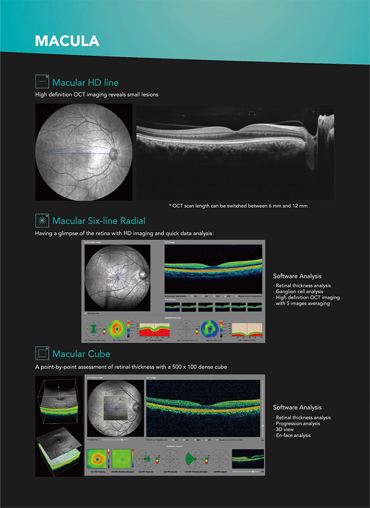
| Scan patterns | Macular :HD line scan(6mm/12mm),3D scan(6mm*6mm), 6-Line radial scan , multi(X-Y:5*5) Disc:3D scan(6mm*6mm) Anterior:HD Line scan: (6/16mm), 6-line radial scan | |
| FUNDUS IMAGING | ||
| Minmum pupil diameter | 3.0mm | |
| Field of view | 40 Degree *30 degree | |
| SOFTWARE ANALYSIS | ||
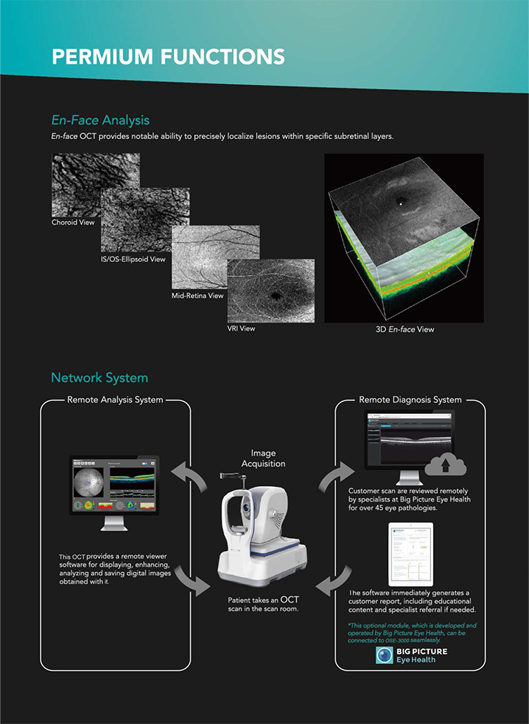
| Macula | Retina thinkness analysis , 3Dview,En-face analysis , Progression analysis, EDI function | |
| Glaucoma | RNFL analysis, Ganglion cell analysis, Cup-disk analysis, Prgoression analysis, OU comparative analysis | |
| Anterior segment | Progression Corneal thickness analysis , Epithelial thinckness analysis | |
| Others | DICOM conformance, Remote viewer software available | |
| ELECTRICAL AND PHYSICAL | ||
| weight | 28.8kg | |
| Dimension | 532mm(l)*360mm(W)*540MM(H) | |
| Source voltage | AC 100-240V | |
| Frequency | 50Hz-60Hz | |
| Power input | 180VA | |